

Cranial cruciate injury is the most common orthopedic injury suffered by dogs of all sizes and breeds. This is usually apparent to owners as a sudden rear leg limp that occurs after some type of activity. This injury is similar to an “ACL” tear in humans. However, the anatomy of the dog differs in the fact that this ligament is utilized to a greater extent including activity and passive stance. With an injured cruciate ligament the dog’s knee joint becomes very unstable, often painful, and may lead to injuries of the meniscus (cushion structure) and cartilage tissues. Most patients require a surgical procedure to stabilize the stifle (knee) joint. Riverview Veterinary Clinic performs two techniques for this injury: the lateral suture and the Tibial Tuberosity Advancement (TTA-2). The decision between which repair would be appropriate for your pet can be made at a consultation with Dr. Marion. Factors such as breed, size, age, activity level, and owner expectations may indicate a preferred procedure.
Lateral Suture
The lateral suture method involves placing a nylon material on the outside of the knee in a similar orientation to the injured cranial cruciate ligament. This stabilizes the joint and after the nylon degrades (weeks to months) fibrous tissue provides long term stabilization. The majority of dogs recover 80-85% of normal within 6 months and may improve further by 1 year. However, smaller dogs are better candidates for this procedure compared to larger dogs, active dogs, or working dogs.
Tibial Tuberosity Advancement (TTA, TTA-2)
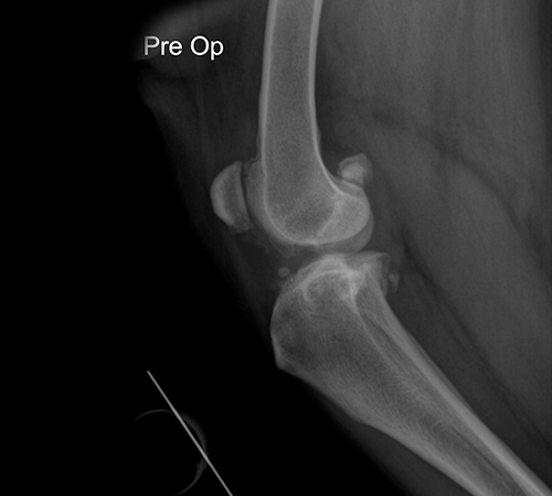
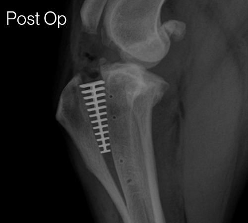
The Tibial Tuberosity Advancement (TTA) technique was developed in 2004 in efforts to improve the outcome of surgically repaired cruciate injuries and reduce the severity of osteoarthritis that follows. This procedure changes the mechanics of the knee joint to compensate for the deficient cruciate ligament. A “cage” and plate secured with forks and screws are used to advance the tibial tuberosity (and therefore the insertion of the patella ligament). Compared to other repair methods, the TTA offers a lower complication rate and excellent recovery. Typical recovery for patients undergoing this procedure is limited activity for 8-10 weeks, but most are full weight bearing within 2 weeks. Patients receiving this repair have a good to excellent prognosis for returning to full athletic activity. Additionally, associated osteoarthritis formation within the knee joint is lower with the TTA compared to other repair techniques. Dogs of all sizes, breeds, and ages can benefit from the TTA or TTA-2.
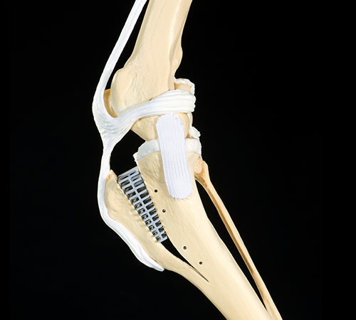
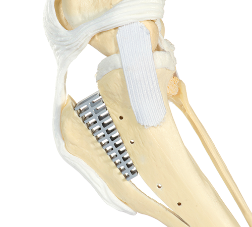
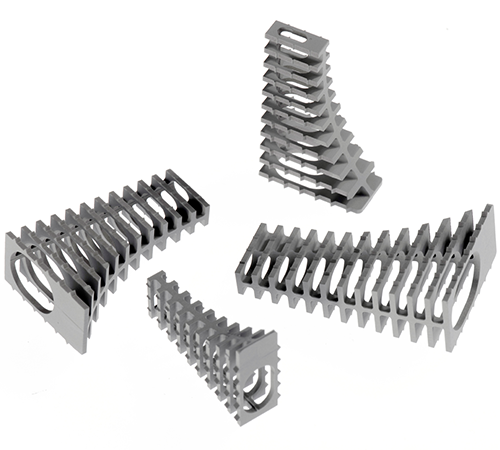
The TTA-2 is the next improvement of this procedure that uses less implant material and preserves more of the surrounding soft tissue compared to the TTA. The TTA-2 consists of placing a single implant made of coated titanium within the tibial bone that advances the insertion of the patella tendon forward. The implant cage is coated with a ceramic material that stimulates bone growth to accelerate healing. A plate and fork is unnecessary with the TTA-2. This achieves the change in biomechanics necessary to stabilize the joint and reduce cartilage damage while preserving more of the surrounding soft tissue due to the minimally invasive approach.
Benefits of TTA-2
- Rapid recovery: full weight bearing within 2 weeks for most cases
- Good prognosis for return to full activity and reduction of lameness
- Decreased progression of osteoarthritis
- Less invasive than TTA, TPLO
- Lower complication rate
- Additional Information
- Images courtesy of KYON Pharma, Inc.
Collection of tissue sample for stem cell banking can also be performed during a TTA-2 repair. While the TTA-2 has the potential to minimize osteoarthritis progression, it will likely still occur. Stem cells have been used to decrease the pain and increase mobility associated with osteoarthritis. The stem cells are harvested now and banked for future use towards any future osteoarthritis (or other conditions) that may develop in the future. For more information on stem cells click here.